MRI Tables
MRI scan (also MRI scan or Magnetic Resonance Imaging (MRI)) is a technique in which radio waves and magnetism are typically used to form images of the body's interior.
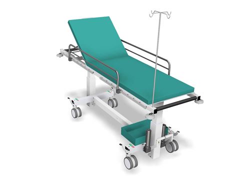
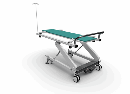
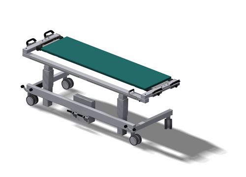
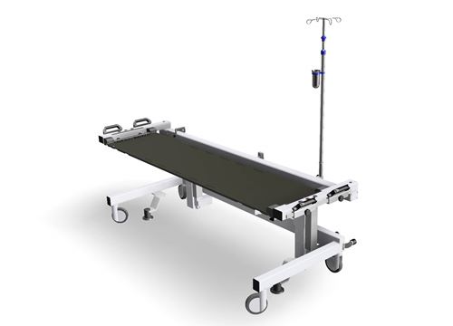
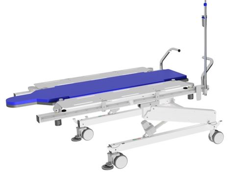
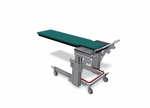
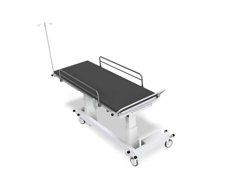
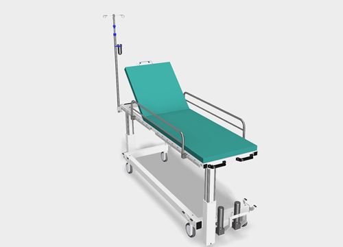
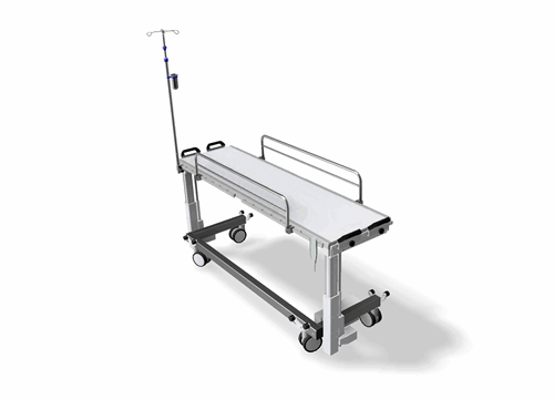
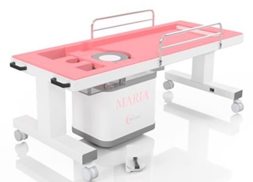
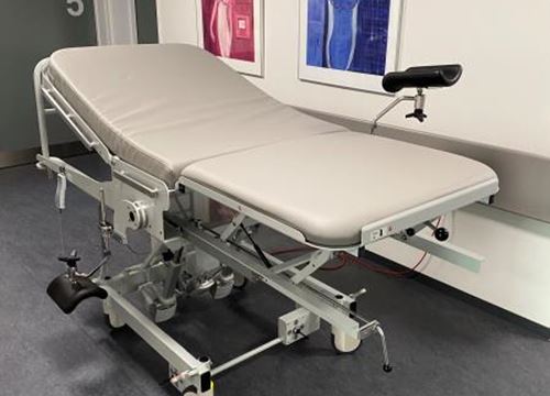
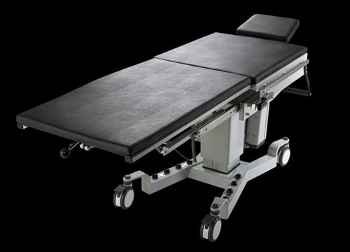
The table is used to drive a patient into the MRI Scanner to slide the patient or lift the patient onto the Scanner table. All materials used must be antimagnetic, hydraulic pump and cylinders are always used for lifting movement.
CT Tables
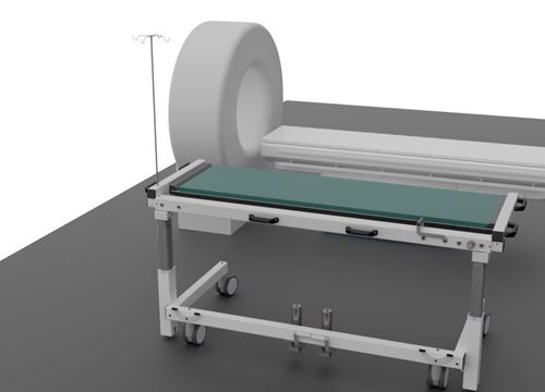
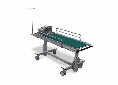
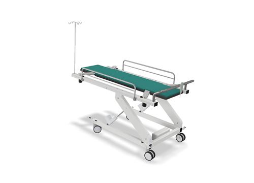
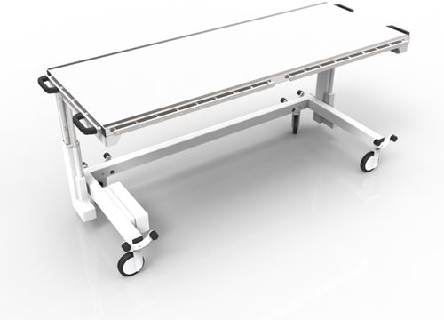
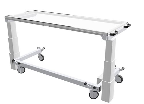
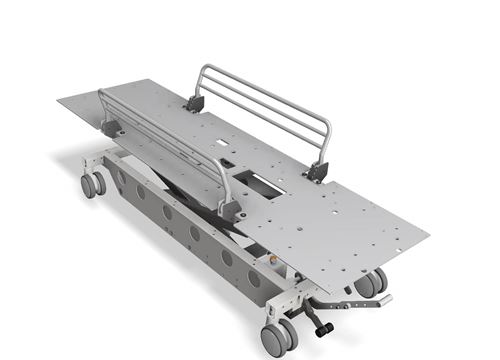
CT Tables are tables which is used to bring the patient from the preparation room or X-ray room to the CT Scanner and lift or sliding the transfer board with the patient up on CT Scanner table for X-ray scanning.
CT scan
CT scan (computer tomography) is an X-ray examination that provides very detailed images of the body's internal organs.
A CT scanner forms sectional images of the body using an X-ray tube that rotates around a bearing running through the scanner. A CT scan collects a very large number of measurement data. These measurements depend on how much the different types of tissue absorb.
Since different tissues absorb X-rays to varying degrees, the computer can then calculate cross-sectional images of the body's interior and display them on a screen.
A CT scanner is suitable for examining:
Size and location of the cancerous nodule as well as the nodule's relations to the surrounding tissue
Acute injuries e.g. bone injuries and internal bleeding
The lungs
Calcifications, e.g. in the urinary tract
Certain bone changes e.g. bone tumors
Acute bleeding
X-ray Tables
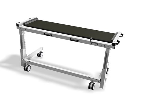
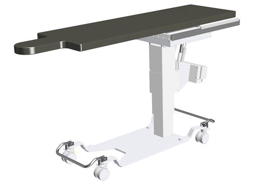
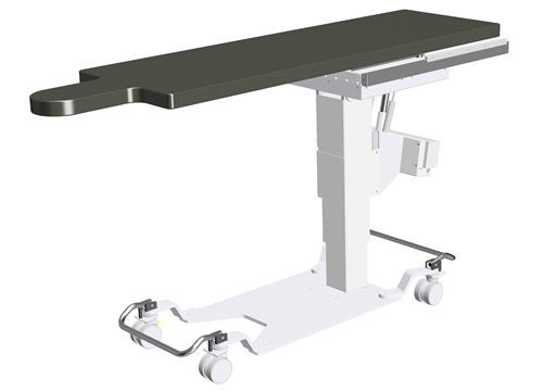
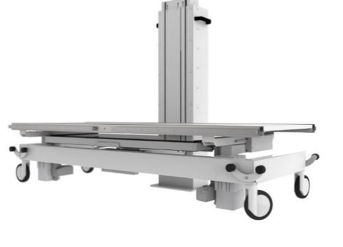
Our X-ray tables can be used for most X-ray examinations as all our X-ray tables have X-ray transparent tabletops.
Ordinary X-ray examinations
Ordinary X-ray examinations are used, for example, to examine the skeleton and chest. When examining smaller bones, a very small dose of X-rays is used.
Transillumination studies
During X-ray examinations, the X-rays pass through the body and can then be seen on a screen as "live images". Transillumination is used e.g. for examinations of the esophagus, stomach and intestinal tract as well as the blood vessels, where you can simultaneously follow some contrast agent.
Stitching & Scoliosis
Scoliosis is one or more deformities in the spine - often or almost always full of a degree of rotation between the individual vertebrae - a so-called torsion.
Stitching unit intent of use:
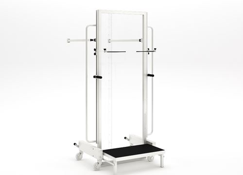
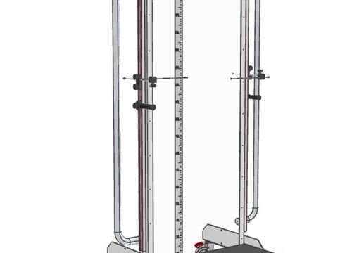
Stitching unit is used for examination of Scoliosis and as the name implies, a stitching of X-Ray images.
When stitching, you take several X-Rays down along the back and put them together on a PC screen, they say you sew them together.
Scoliosis unit intent of use:
Scoliosis unit is used for examination when:
Scoliosis is the term for a type of skew in which the spine is S-shaped or C-shaped when viewed from behind
Customer solutions
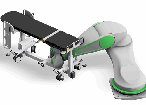
Customer solutions usually start with a contact from a customer who wants a special solution, we then have some initial meetings with the customer, where we find a solution for the customer. We then make a requirements specification together with the customer which is the start of the production of the prototype. We work closely with the customer during the prototype production and after the completion of the prototype, there is a reassessment before the start of the final product.
PCNL Tables
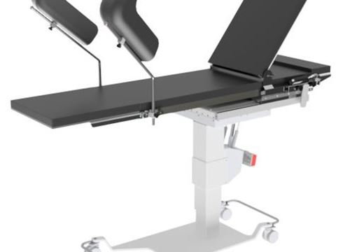
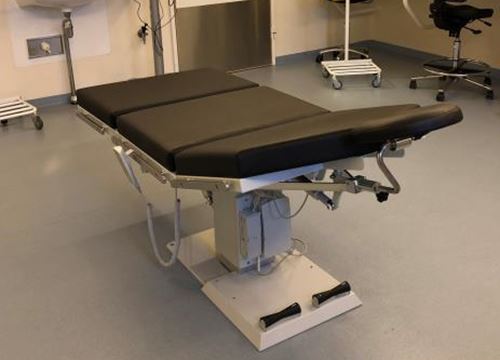
Our PCNL Table with X-ray transparent tabletop, is special designed for kidney stone surgery, with continuous X-ray overview.
Kidney stone surgery
The surgery is performed by making a small 1 cm incision in the patient’s flank area. A tube is placed through the incision into the kidney under x-ray guidance. A small telescope is then passed through the tube to visualize the stone, break it up and remove it from the body. If necessary, a laser or other device called a lithotripter may be used to break up the stone before it can be removed. This procedure has resulted in significantly less post-operative pain, a shorter hospital stays, and earlier return to work and daily activities when compared to open stone surgery.
Transillumination studies
During X-ray examinations, the X-rays pass through the body and can then be seen on a screen as "live images". Transillumination is used e.g. for examinations of the esophagus, stomach and intestinal tract as well as the blood vessels, where you can simultaneously follow some contrast agent.

